Embarking on a miraculous journey, the stages of a fertilized chicken egg take us on a fascinating exploration of life’s beginnings. Every step holds wonder and awe, from the blastoderm’s delicate formation to the organs’ intricate development. Starting with the mesmerizing fertilization process, where the sperm meets the Egg, a remarkable sequence unfolds. The Egg then progresses through cleavage, gastrulation, and neurulation, giving rise to the embryo’s major structures.

Stages of a Fertilized Chicken Egg
Fertilization: During fertilization, a sperm cell from the rooster combines with the egg cell in the hen’s oviduct. This fusion creates a zygote containing the complete genetic information required for development.
Cleavage: The zygote undergoes rapid cell division, known as cleavage. This process produces a cluster of smaller cells called blastomeres. Cleavage continues, forming a hollow sphere known as a blastula.
Gastrulation: Gastrulation is a critical stage where the blastula transforms into a three-layered structure called the gastrula. The three layers are ectoderm, mesoderm, and endoderm, giving rise to different tissues and organs.
Neurulation: Neurulation is the formation of the neural tube, which will form into the brain and spinal cord. The ectoderm layer thickens and folds, creating the neural groove and eventually closing to form the neural tube.
Organogenesis: During organogenesis, the embryo’s organs and systems develop. The heart, liver, kidneys, and vital organs take shape, and limb buds appear, which will develop into wings or legs.
Growth and Maturation: The embryo continues to grow and mature, developing feathers, beaks, and other distinguishing features. The yolk sac, gives nutrients to the developing embryo, shrinks as the chick’s internal structures become self-sustaining.
Embryonic Development of a Fertilized Chicken Egg
The embryonic development of a fertilized chicken egg is a complex process. After fertilization, the Egg changes carefully orchestrated events of changes that lead to the formation of a fully developed chick. Within the first 24 hours, the Egg undergoes cleavage, a process of rapid cell division.
By day 3, the embryo forms a primitive streak, which gives rise to the central nervous system. Over the next few days, the major organ systems, including the heart, digestive system, and limbs, begin to develop. By day 21, the chick is ready to hatch, using an egg tooth to crack the shell and emerge into the world.
Chicken Egg Fertilization Process and Stages
The process of chicken egg fertilization is an intricate sequence of events. When a rooster mates with a hen, the rooster’s sperm is transferred to the hen’s reproductive tract. Once inside, the sperm cells travel to the oviduct, where they can fertilize the Egg. Fertilization occurs when a sperm cell penetrates the Egg and fuses with its nucleus, forming a zygote. The fertilized Egg then begins its developmental journey.
The zygote undergoes cell division in the first 24 hours, creating multiple cells. By day 3, a structure called the blastodisc forms, which contains the embryo’s genetic material. Over the next few days, the embryo develops key structures such as the neural tube, heart, and digestive system.
Understanding the Growth Stages of a Fertilized Chicken Egg
From conception to hatching, the Egg goes through distinct phases. Cell division occurs rapidly, forming the primitive streak and developing the embryo’s backbone and nervous system. As the days progress, major organs, limbs, and features like the beak and eyes begin to form. Blood circulation begins, and feathers develop. By the end of incubation, the chick is fully formed and ready to hatch. Each stage showcases the intricate development process, from the initial cell division to the emergence of a vibrant, living chick.
In case you missed it: The Best Way to Keep Chickens Laying Eggs During the Winter

Detailed Timeline of a Fertilized Chicken Egg’s Development
- Day 1: Within the first 24 hours, the primitive streak forms, initiating the development of the embryo’s head, backbone, and nervous system. The alimentary tract and blood islands also begin to appear.
- Day 2: Blood islands start linking and forming the vascular system, while the heart forms separately. By the 44th hour, the heart and vascular system merge, and the ear starts to form.
- Day 3: The embryo’s position allows better visibility. The heart begins to beat, blood circulation starts, limb buds form, the nose develops, and blood vessels thicken. The allantois and vitelline membranes also begin to form.
- Day 4: The amniotic cavity forms, protecting and allowing movement for the embryo. The tongue develops, the allantoic vesicle appears, and the brain divides into four parts. The eyes start to form.
- Day 5: The embryo takes on a distinct C shape, and sex differentiation occurs. Reproductive organs begin to form.
- Day 6: The beak and vitelline membrane continue to grow, fissure form between fingers, and the eyes, heart, and brain become more prominent. The voluntary movement begins.
- Day 7: The neck separates the head from the body, the beak enlarges, the brain settles, and the comb develops.
- Day 8: Upper and lower beaks, legs, and wings differentiate. The neck elongates, the brain settles into its cavity, and the external ear opening forms.
- Day 9: Claws appear, the allantois grows larger, vascularization increases, and feather follicles start to develop.
- Day 10: The beak hardens, the egg tooth appears, nostrils form, eyelids grow, limb portions lengthen, and feather follicles cover limbs.
- Day 11: Eyelids enlarge, the allantois reaches maximum size, the vitellus begins to shrink, and tail feathers appear.
- Day 12: Feather follicles surround the ear, eyelids continue to grow, and toes fully form.
- Day 13: The allantois merges with the chorion, scales on the legs, and toenails form.
- Day 14: Down covers the entire body and the chick positions itself for hatching.
- Day 15: Down and embryo continue to grow, vitellus absorption accelerates, and the chick moves into the pipping position.
- Day 16: The embryo assumes the pipping position, absorbs albumen, and Egg darkens.
- Day 17: The renal system produces urates, the beak points towards the air cell, and albumen absorption completes.
- Day 18: “Lockdown” day, where turning stops, humidity increases and vitellus absorption begins.
- Day 19: Vitellus absorption accelerates, and the beak aligns with the air cell, preparing for the internal pip.
- Day 20: Internal pip occurs as the chick breaks the internal membrane and breathes air. Vitellus absorption completes, and external pipping may occur.
- Day 21: The chick starts to break the shell, known as zipping, and eventually hatches, ready for the brooder.
Stages of Embryogenesis in a Fertilized Chicken Egg
- Before Egg Laying: Fertilization occurs, and living cells divide and grow. Cells segregate into groups with specific functions.
- Between Laying and Incubation: No growth; the embryo remains inactive.
- During Incubation: A resemblance to a chick embryo appears. Alimentary tract and vertebral column start forming. Nervous system and head development begin. Eyes become visible.
The fertilized Egg undergoes cell division and differentiation during these stages, forming vital organs and body structures. The embryo’s appearance gradually changes with the development of the head, backbone, limbs, beak, feathers, and reproductive organs. As incubation progresses, the embryo prepares for hatching, positioning itself and absorbing the yolk sac.
Cell Division and Differentiation in a Fertilized Chicken Egg
Cell division in a fertilized chicken egg is crucial for growth and development. The Egg forms a blastodisc, which contains genetic material for the embryo’s development. As cell division progresses, it transforms into a blastoderm, which forms various tissues and organs. Cell differentiation occurs, with some cells becoming ectoderm, endoderm, and mesoderm cells.
These processes are tightly regulated, ensuring the proper development of the embryo. Understanding cell division and differentiation in fertilized chicken eggs offers insights into embryonic development, with implications for developmental biology and regenerative medicine.
Morphological Changes During the Development of a Fertilized Chicken Egg
During the development of a fertilized chicken egg, morphological changes occur. Initially, the Egg consists of a single cell, which undergoes rapid cell division. Over time, distinct structures begin to form, including the primitive streak, head, backbone, limbs, beak, and feathers.
As development progresses, the chick’s organs and body systems take shape, such as the digestive, respiratory, and circulatory systems. The embryo’s appearance becomes increasingly bird-like, with scales, claws, and a hardened beak. These morphological changes culminate in chick hatching after approximately three weeks of incubation.
In case you missed it: 10 Best Chicken Breeds For Producing Large Brown Eggs

Hatching Process and Stages of a Fertilized Chicken Egg
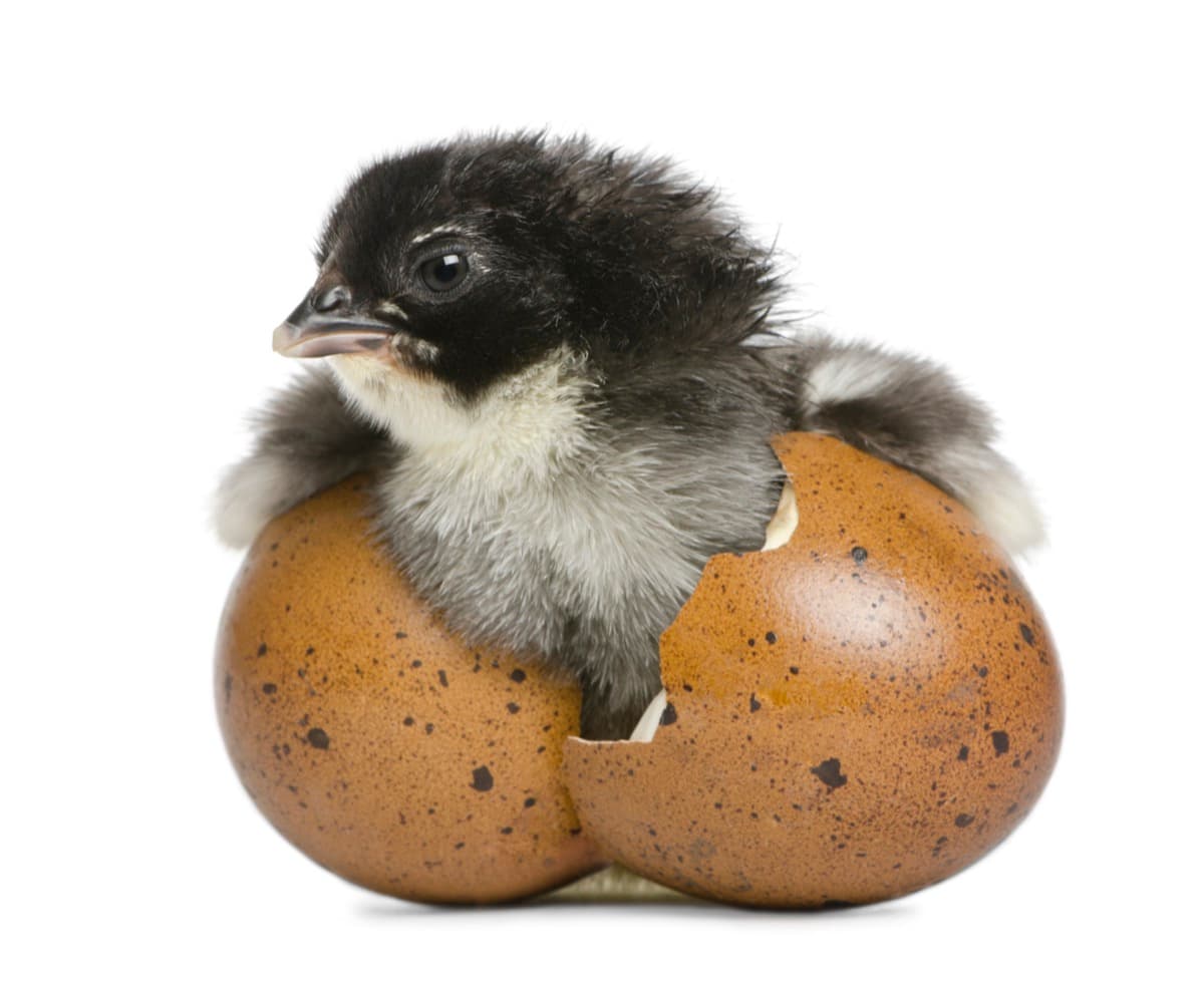
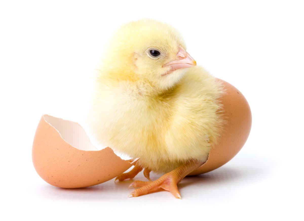
The hatching process of a fertilized chicken egg involves distinct stages. First, the chick pierces the air cell with its beak, initiating pulmonary respiration. It then cuts through the shell using the egg tooth and neck muscles. After breaking free, the chick rests, allowing the navel openings to heal and it’s down to dry. Gradually, it gains strength, walks, and adapts to its new environment. The process usually takes 24-48 hours from the initial pip to complete hatching.
Conclusion
The journey of a fertilized chicken egg from conception to hatching is a unique and intricate process. Through stages of development, the embryo transforms, forming vital organs and structures. Finally, the chick breaks free from the shell, ready to begin its new life.
- Feed Your Flock for Less: Top 10 Tips to Save on Chicken Feed
- Ultimate Guide to Ossabaw Island Hog: Breeding, Raising, Diet, and Care
- Hatching Answers: The Top 10 Reasons Your Chickens Aren’t Laying Eggs
- Eggs and Economics: Breaking Down the Cost of Raising Backyard Chickens
- Defend Your Greens: Proven Methods to Keep Iguanas Out of Your Garden
- Ultimate Guide to Cinnamon Queen Chicken: A Comprehensive Guide for Beginners
- Ultimate Guide to California Tan Chicken: Breeding, Raising, Diet, Egg-Production and Care
- Ultimate Guide to Marsh Daisy Chicken: Breeding, Raising, Diet, and Care
- 10 Types of Chicken Farming Businesses You Can Start for Profits